trading as Calder Hall Medical Centre
X-Rays (Medical Radiography)
To make an appointment, please call our Reception Desk ![]() (868) 660 7873 or
(868) 660 7873 or ![]() (868) 639 8516
(868) 639 8516
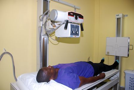
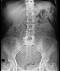
Medical Radiography is a broad term that covers several types of studies that require the visualisation of the internal parts of the body using x-ray techniques. For the purposes of this page radiography means a technique for generating and recording an x-ray pattern for the purpose of providing the user with a static image or images after termination of the exposure. It is differentiated from fluoroscopy, mammography, and computed tomography. Radiography may also be used during the planning of radiation therapy treatment. It is used to diagnose or treat patients by recording images of the internal structure of the body to assess the presence or absence of disease, foreign objects, and structural damage or anomaly. During a radiographic procedure, an x-ray beam is passed through the body. A portion of the x-rays is absorbed or scattered by the internal structure and the remaining x-ray pattern is transmitted to a detector so that an image may be recorded for later evaluation. The recoding of the pattern may occur on film or through electronic means. When are X-Rays Used?Radiography is used in many types of examinations and procedures where a record of a static image is desired. Some examples include
Women who are pregnant should inform their doctor.
|
|
© 2016 Specialist Clinics of Tobago Ltd.- All rights reserved -- Last updated: November 28, 2017

